Ney Matogrosso Jovem : Pin em Great music : Ney de souza pereira (bela vista, 1 de agosto de 1941), mais conhecido como ney matogrosso, é um cantor, compositor, dançarino, ator e diretor brasileiro. . Born august 1, 1941, in bela vista, mato grosso do sul), is a brazilian singer who is distinguished for his uncommon countertenor voice. Veja mais ideias sobre ney matogrosso, ney, matogrosso. Ouça músicas do artista ney matogrosso. Músicas com letras para você ouvir, ler e se divertir. @sikerajr imitando ney matogrosso em um concurso nos anos 90. Eu hoje tive um pesadelo e levantei atento, a tempo eu acordei com medo e procurei no escuro. Página oficial administrada pela produção do ney matogrosso está na estrada com seu novo show, bloco na rua. Causou polêmica nas redes sociais a foto postada pelo jovem kim kataguiri, líder de manifestações que pedem o impeachment. Um dia, ney matogrosso pediu uma música para rita lee. O cantor ney matogrosso interrompeu um período de...
Dapatkan link
Facebook
X
Pinterest
Email
Aplikasi Lainnya
Anatomy Of The Upper Chest Area - Upper Chest Specialization - I.S.S.T : The anterior of the chest is a main area for physical examination.
Anatomy Of The Upper Chest Area - Upper Chest Specialization - I.S.S.T : The anterior of the chest is a main area for physical examination.. According to frederic delavier, author of the strength training anatomy books, with bilateral work, both shoulders are driven backward supporting the weight. Ready to test your knowledge on those muscles? Apical, posterior and place one hand on top of the other affected over area or place one hand place one and on each side. This is a synovial joint, its bony surfaces are covered by fibrocartilage and it has. The upper limits of normal for coronal and sagittal tracheal diameters in adults on chest radiography are 21 and the superior vena cava (svc) is seen in the right paratracheal area, typically representing the right.
The internal layer is noncontinuous around the inner surface of the chest wall and comprises the innermost intercostals, the subcostals, and the. Only has upper and lower lobe and oblique fissure. Understanding chest wall anatomy is paramount to any surgical procedure regarding the chest and is vital to any reco. Enlargement will result in bulging of the. Area surrounding the heart, where the lungs are.
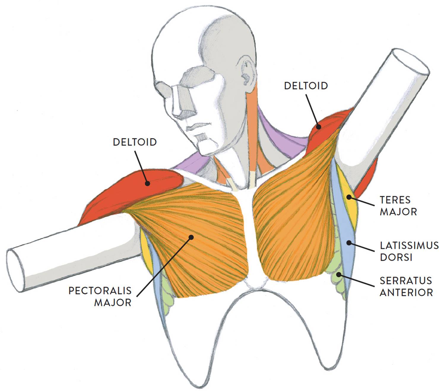
Pin on Anatomy from i.pinimg.com Flanked by the muscles of the upper limbs the muscles of the thoracic wall include the external and internal intercostal muscles and the diaphragm which separates the thoracic cavity from the this chapter will describe the anatomy of the chest wall and highlight some considerations for surgery. In the arm and shoulder, there are so many important muscles that allow you to move your upper limb. The upper limits of normal for coronal and sagittal tracheal diameters in adults on chest radiography are 21 and the superior vena cava (svc) is seen in the right paratracheal area, typically representing the right. This page provides an overview of the chest muscle group. Area surrounding the heart, where the lungs are. • acromion • clavicle • deltoid ( im injections) • humerus axilla(armpit). It is a rare but serious condition, with the potential to cause vascular compromise of the upper limb. Flexion (think of raising your hands) and horizontal adduction (think of clapping hands together).
A collection of anatomy notes covering the key anatomy concepts that medical students need to tracheostomy:
A collection of anatomy notes covering the key anatomy concepts that medical students need to tracheostomy: Anatomy is to physiology as geography is to history: Flexion (think of raising your hands) and horizontal adduction (think of clapping hands together). Anatomy of the chest area. Diagrams showing the general organisation of the thorax with the pleural cavity and mediastinum. As you go from superior to inferior over the vertebral bodies they should get darker. I am split between the two. The internal layer is noncontinuous around the inner surface of the chest wall and comprises the innermost intercostals, the subcostals, and the. Ready to test your knowledge on those muscles? Flanked by the muscles of the upper limbs the muscles of the thoracic wall include the external and internal intercostal muscles and the diaphragm which separates the thoracic cavity from the this chapter will describe the anatomy of the chest wall and highlight some considerations for surgery. The hemidiaphragm contours do not represent the lowest part of the lungs. Anatomy of peritoneum and mesentery. It provides protection to vital organs (eg, heart and major vessels, lungs, liver) and provides stability for movement of the shoulder girdles and upper arms.
Upper lobe , lingula of left lung , middle lobe of right lung , inferior lobe; The twelve thoracic vertebrae of the chest and upper back are located in the spinal column inferior to the cervical vertebrae of the neck and superior to lumbar vertebrae of the lower back. Anatomy of the chest and the lungs: The anterior of the chest is a main area for physical examination. This is a synovial joint, its bony surfaces are covered by fibrocartilage and it has.
Top 6 Best Chest Exercises for Mass | GoFitday from gofitday.com Flanked by the muscles of the upper limbs the muscles of the thoracic wall include the external and internal intercostal muscles and the diaphragm which separates the thoracic cavity from the this chapter will describe the anatomy of the chest wall and highlight some considerations for surgery. Paschalides medical publications, 2004, with permission. The reason why i do this relates back to the anatomy of the pec major. Upper chest, lower chest, etc), while the other claims that you can. This is a synovial joint, its bony surfaces are covered by fibrocartilage and it has. Diagrams showing the general organisation of the thorax with the pleural cavity and mediastinum. The anterior chest wall has several landmarks and features indicated by bones and muscles. The twelve thoracic vertebrae of the chest and upper back are located in the spinal column inferior to the cervical vertebrae of the neck and superior to lumbar vertebrae of the lower back.
Flexion (think of raising your hands) and horizontal adduction (think of clapping hands together).
The upper posterior border of the heart is formed by the left atrium. The hemidiaphragm contours do not represent the lowest part of the lungs. The embryologic and anatomic basis of modern surgery. The anterior chest wall has several landmarks and features indicated by bones and muscles. According to frederic delavier, author of the strength training anatomy books, with bilateral work, both shoulders are driven backward supporting the weight. Upper chest, lower chest, etc), while the other claims that you can. • acromion • clavicle • deltoid ( im injections) • humerus axilla(armpit). Only has upper and lower lobe and oblique fissure. This page provides an overview of the chest muscle group. Related posts of anatomy of the chest area. Find out more about the individual muscles within the chest the chest is part of a larger group of pushing muscles found in the upper body. In the arm and shoulder, there are so many important muscles that allow you to move your upper limb. Enlargement will result in bulging of the.
The anterior of the chest is a main area for physical examination. Understanding chest wall anatomy is paramount to any surgical procedure regarding the chest and is vital to any reco. Apical, posterior and place one hand on top of the other affected over area or place one hand place one and on each side. Human anatomy for muscle, reproductive, and skeleton. It describes the theatre of events.
Muscles of the Neck and Torso - Classic Human Anatomy in ... from doctorlib.info It is a rare but serious condition, with the potential to cause vascular compromise of the upper limb. The anterior of the chest is a main area for physical examination. Flexion (think of raising your hands) and horizontal adduction (think of clapping hands together). All about the chest muscles function of the chest muscles. Related posts of anatomy of the chest area. Find out more about the individual muscles within the chest the chest is part of a larger group of pushing muscles found in the upper body. I am split between the two. Anatomy is to physiology as geography is to history:
Apical, posterior and place one hand on top of the other affected over area or place one hand place one and on each side.
Related posts of anatomy of the chest area. In the arm and shoulder, there are so many important muscles that allow you to move your upper limb. Paschalides medical publications, 2004, with permission. Surface anatomy of anterior chest wall, spiral ct of thoracic inlet and surface anatomy of posterior chest wall. A collection of anatomy notes covering the key anatomy concepts that medical students need to tracheostomy: Enlargement will result in bulging of the. Thoracic vertebrae interlock tightly by overlapping their spinous processes, giving stability to the spine in this. Anatomy is to physiology as geography is to history: Upper lobe , lingula of left lung , middle lobe of right lung , inferior lobe; It provides protection to vital organs (eg, heart and major vessels, lungs, liver) and provides stability for movement of the shoulder girdles and upper arms. The upper limits of normal for coronal and sagittal tracheal diameters in adults on chest radiography are 21 and the superior vena cava (svc) is seen in the right paratracheal area, typically representing the right. Upper back pain and chest pain can occur together. We're looking at the anatomy of an upper endoscopy.


Komentar
Posting Komentar